Breast Cysts
Breast cysts make up 15% of all breast lumps. They are fluid filled lumps in the breast. The incidence of breast cysts increases up until menopause. They can be multiple and occur in both breasts. Cysts can appear suddenly. They are influenced by female hormones and can fluctuate in size. They are not associated with an increased risk of breast cancer. Sometimes management includes aspiration of the cyst.
A complex cyst is one that has an unusual appearance on ultrasound. It may have a solid component or a septation (band of tissue within it). A complex cyst is usually investigated with aspiration or biopsy. Occasionally it may require surgery.
Fibroadenomas
Fibroadenomas are benign tumours of the breast and the most common cause of a breast lump in young women. In fact they can occur in up to 9% of women.
In a fibroadenoma there is overgrowth of the cells lining the milk ducts of the breast and of the supporting tissue in between the ducts. This overgrowth produces a benign lump.
Fibroadenomas contain breast cells and like anywhere else in the breast can develop cancer. The risk of developing a cancer in a fibroadenoma is no more than elsewhere in the breast.
Fibroadenomas can vary in response to normal cyclical hormone fluctuations.
Management of fibroadenomas usually involves a biopsy to confirm the diagnosis and then observation. Excision may be performed if there are any doubts as to the nature of the lump, if it is large, if it is increasing in size or if it is causing symptoms. It may be a patient’s preference to have a fibroadenoma excised.
Phyllodes Tumours
It can be sometimes be difficult to differentiate between a fibroadenoma and another type of breast tumour called a phyllodes tumour. The term phyllodes comes from the Greek, meaning leaf like, in reference to their appearance on histologic examination. Phyllodes tumours can be benign, borderline or malignant. If a fibroadenoma is found to be enlarging or atypical on biopsy, excision is usually recommended in order to exclude a phyllodes tumour. The vast majority of phyllodes tumours are benign and cured with excision. Borderline and malignant phyllodes are rare and require excision with a margin, regular follow up and occasionally other treatment.
Atypical Lesions found during Breast Screening
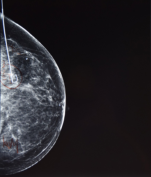
Breast screening occasionally identifies areas of concern such as calcification on mammogram or a lesion on ultrasound. These are usually assessed with needle biopsy and if they aren’t definitely diagnosed as benign may require surgical excision. As these lesions can’t be felt, they require a procedure called hookwire localisation immediately prior to surgery. The hookwire is placed in the breast imaging department using local anaesthetic and ultrasound or mammogram to guide the wire into the correct location. Your surgeon will use the wire as a guide to know which area of breast tissue needs to be excised, during your surgical procedure later that day.

